Checking array microscopes with ramona optics
ReadAt Argolight, we believe that the real proof of a product’s impact comes from the people who use it every day. That’s why we invite our satisfied users to share their stories.
Aurélien Bègue, R&D director at Ramona Optics, explains how Argolight’s technology has brought tangible benefits to their workflow for the Vireo system, a high-throughput, multi-camera array microscope. Here is Ramona Optics’ story, which walks through the evolution of this partnership, highlights the unique features of the Vireo, and explores how a company delivers repeatable, high-quality results for users around the globe.
Aurélien holds a PhD in Neurosciences and Applied Optics and previously served as an Imaging Core Scientific Director at Harvard Medical School. His academic research focused on developing new microscopy tools for optogenetic experiments using wavefront shaping techniques. He has co-authored multiple publications using Ramona’s MCAM technology.
What we do
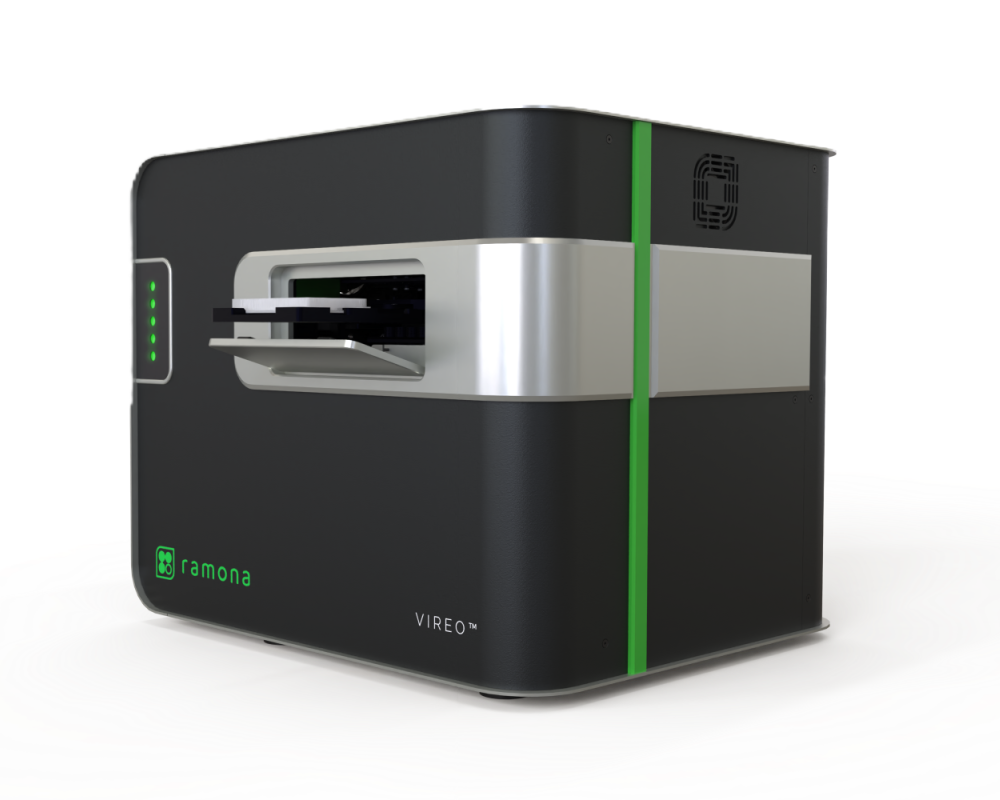
The Vireo is the second product at Ramona. We naturally evolved from imaging larger model organisms at lower resolution to imaging cellular samples at higher resolution in multiwell plates. We developed the Vireo to be the world’s fastest live-cell imaging system. Not only does our camera array allow for multiplexing the imaging of multiple wells simultaneously and thus increasing throughput, but it also allows for observation of live events across multiple conditions at video frame rate.
The arraying of multiple objectives makes a difference from other microscopy systems. It enables live imaging over 24 wells simultaneously or to quickly scan an entire plate within a minute within automated workcells.
Maybe more surprising is our ability to Z-stack large volumes. This means a user doesn’t rely on an autofocus mechanism or go through a long pre-step of creating a focus map. The result: dozens of samples imaged at once, all in focus and delivering analyses in a few minutes after the acquisition ends.
Bringing QC into the project
I think microscope users are demanding access to the newest technology, the easiest interaction possible to get the prettiest image that will generate the best graphs for their manuscripts.
With the multiplication of techniques and post-processing tools at the user’s disposal, it is very important to be clear about what stays within the boundaries of quality imaging. We are integrating machine learning into our analysis tools, and it’s essential that we provide accurate analysis, which starts with high-quality imaging. In image analysis, the old rule of “you cannot expect good results from bad images” imposes that the microscope builders ensure the upstream quality.
Argolight contacted us about a year and a half ago, explaining the benefits of Fluorescence QC. I was definitely intrigued on two counts. First, our systems inherently rely on multiple objectives, and ensuring uniformity among them is crucial. Second, as a French person myself, it was particularly exciting to receive this proposal from a French company that appears to be advancing cutting-edge technology.
“In image analysis, the old rule of “you cannot expect good results from bad images” imposes that the microscope builders ensure the upstream quality.”
Developing the right tool
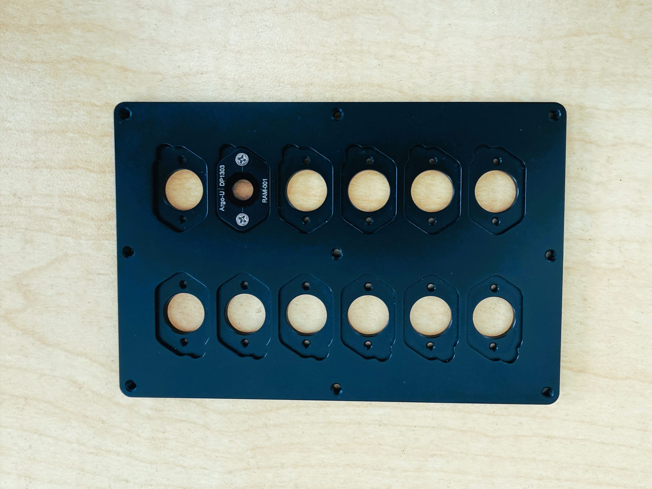
We use a custom version of the Argo-LM slide. Before we landed on this format, we tried multiple formats and even the HM slide.
We are still in the early part of the process, but I hope to equip the field support team with a tool to check systems they visit and track them over time. Throughout the process, the Argolight team supported us on the imaging pattern and mechanical designs. The close collaboration really made this process a breeze for us.
In our assembly process, we are using the Argo-tile during the calibration step. It serves both as a readout of the quality of the system and as a way to fine-tune parameters for the different components of the array. Ensuring the uniformity across the array is definitely the primary goal.
The next steps
Being able to guarantee consistent and reliable results over the lifespan of our instruments is the grail. Many components have different lifetimes and are solicited to different extents. The only way to monitor and keep the performance is to keep up with the maintenance and perform QC at regular intervals.
By weaving Argolight’s proven QC tools into the flexible Vireo system, Ramona delivers a brighter, more dependable illumination platform that meets the rigorous demands of modern microscopy.
Ramona Optics is setting a new standard for data-rich experimentation with its revolutionary multi-camera array microscope (MCAM), a high-throughput, real-time 4D imaging technology that continuously captures intricate cellular dynamics across multiple conditions at scale. MCAM’s efficient, cost-effective imaging enables researchers to observe cellular behavior in unprecedented detail.
I3S
Microscopy is at the heart of countless biomedical discoveries—but how do you make sure the images you produce today are as reliable as those from...
User case: Kyoto University x Argolight – Fluorescent proteins observation
Matsuda Lab, Graduate School of Biostudies/Graduate School of Medicine, Kyoto University Introducing Argolight slides - A New Quality Control...
Quality control of HCS-HTS fluorescence imaging systems
In the landscape of high-content screening (HCS) and high-throughput screening (HTS) fluorescence imaging systems, precision and reliability take...
