We have interviewed one of our first customers, who works in pharmaceutical diagnostics. He provided great insights into some of the concerns in the field. We will call him John (not his/her real name). This article is voluntarily anonymous to respect the strict endorsement rules of our customer’s institution.
We virtually sat with him one morning to exchange about his work and experience with Argolight.
Good morning John, can you tell us what is your job and field of expertise?
Hello Argolight! My work is about diagnostic technologies applied to tissues taken out of patients for evaluation. “Tissue diagnostics” for short.
Tissue diagnostics, can you tell us more ?
Sure, there are two ways to approach tissue diagnostics. One is molecular testing and the other is antibody labeling. It uses antibodies to stimulate certain molecules in tissue sections that have been put on microscope slides. They are then stained with markers, to tell where proteins are being expressed.
That is your expertise, right ?
Yes, I develop technologies to image new types of markers on tissue sections, what we call multiplex assays. It means looking at multiple biomarkers in a tissue section, not just one marker. You could do that with bright-field microscopy, of course, using different colors as it absorbs different levels of light. And there is also fluorescence microscopy, which uses a wavelength of light to excite the sample and causes it to emit different wavelengths for detection.
“It used to be human-made but since the past 10 years or so, these information are digitally captured and processed by algorithms.”
What is the crucial step using the antibody labeling approach for tissue diagnostics?
It is the interpretation. It used to be human-made but for the past 10 years or so, this information is now digitally captured and processed by algorithms.
It allows searchers to develop more rapidly and accurately assays that provide valuable information on what’s going to happen to the patient, or which medicine is going to benefit them.
That seems great, so thanks to technology, you are able to get results faster?
In theory, yes, but reliability is extremely important in tissue diagnostics. Markers detection implies a very good calibration of the system to ensure that if markers appear brighter, it is not because some channels are receiving more illumination than others.
What approach did you choose and why?
So we need to remove the bias in our detection, to be able to know that differences of results do come from the sample and not from the instrument.
We need to control for the differences from one microscope to another one. So the approach was to buy microscopes, set them up the same way, with the same hardware, and get the same answer and behavior from them.
“We needed to remove the bias in our detection, to be able to know that differences of results do come from the sample and not from the instrument.”
I guess this is where you found Argolight products?
Yes, we started looking for some sort of a calibration slide that would work across multiple wavelengths. A colleague searched for such a tool and quickly found Argolight. We went right ahead and purchased the first product to test.
I have been looking for 20 years for a tool to calibrate a fluorescent microscope across different channels. There is really nothing good out there that one can use easily. Argolight has a unique product.
“I have been looking for 20 years for a tool to calibrate a fluorescent microscope across different channels.”
Why is calibration such an important issue for you?
In my work, I am primarily concerned about the intensity. If we shine a given amount of light, do we get a given amount out? It is usually important for us because the camera only detects spatial coordinates and intensity. And that is the foundation for all the digital algorithms that operate in our technology.
“If we have a lot of variability in the intensities, it creates problems for us.”
If we have a lot of variability in the intensities, it creates problems for us. We want to calibrate for the intensity on all the channels, not just on one channel, across different microscopes. It is a reproducibility issue.
We wanted to check repeatability too, as a temporal consideration: is our microscope intensity the same day after day, week to week, when we image something? The answer is “probably not” on a research-grade instrument.
But “probably not” is not enough given the scope of applications in tissue diagnostics. That is why we wanted to characterize on a regular basis any changes or drifts that were happening over time. We needed a tool that was exactly the same, or very close to exactly the same that we could use each day.
“Is our microscope intensity the same day after day, week to week, when we image something? The answer is ‘probably not’.”

So, you are looking to monitor for intensity both spatially and in terms of value, are there other aspects you care about in your research?
There are spatial elements too that we want to calibrate for as well. We are digitalizing large areas, so we must evaluate the mechanical repeatability of the mechanisms that move slides around. This is a secondary element, as it can be done in other ways.
You have purchased your first product 7 years ago and since you purchase additional products once every two years or so, how do you use these products? Do you still use the first one?
Yes! We are still using the slide, 7 years after our first purchase, and experimenting ways to use them in our calibration protocols. We had the 1st generation of slides with a different glass formulation. We now own the latest generation with the new formula of glass.
“We are still using the slides, 7 years after our first purchase.”
So how do you use the slides nowadays?
There are all kinds of things that drift in the instruments: the bulb’s age, the light guide age, stacking tolerances… Instrument calibration can be done with sensors, but it’s painstaking, it could take days to calibrate that carefully an instrument, so calibrating a room full of instruments is a real question.
The efficient way is to take a standard, image it on a calibrated microscope, get a map at each channel (fluorescence response curve across the sample) to get a fingerprint of what we should expect from the standard on other instruments. We then put this characterized slide on another instrument and see the differences. That how we know that, for instance, we need to increase the exposure time to get the same results.
The calibration slide provides expected results across wavelengths. The idea would eventually be that it becomes a component on the instrument, to get a self-check instrument, that does this routinely and database the results, so we can track drifts over time.
Have you been using the slide continuously over the past years?
There has been a couple of years where we did not use our calibration slide because the project was paused. These kinds of research projects tend to fluctuate. They are a hot topic and we advance on them, then they get shelved for a while because people are working on other things. And they come back because something has been found and is worth pursuing again. But the slides were still reliable after a multiyear pause.
Of course, as any tool, there are still things that could be improved. The current glass formulation seems to have a much higher fluorescence in the blue side of the spectrum, but less fluorescence in the red area. It would be ideal if we had a lot more consistency across the visible spectrum, in terms of fluorescence yield. We would also like to understand better the short-term response of the slide to illumination.
I would recommend the product. It provides a very consistent result that allows characterizing instrument in a way that would be very difficult to do using other technologies.
“The risk is that people do not know how much variability they are introducing into their activities.”
Which advice would you give to users considering Argolight slides?
The first thing most people look at is the price of the slide, it seems high, but it is worth it. The cost/investment is really quite small compared to the risk you have if you do not use something like this to calibrate an instrument. A lot of people assume they get the same results just because they have the same hardware. Hardware can vary from 20 to 30% between instruments, it is more than what most people realize.
The risk is that people do not know how much variability they are introducing into their activities. It can result in people redoing experiments and never getting to the bottom of where the variability is getting from.
In terms of person-hours, it would amount to a fair amount in a clinical research environment. So data-driven science, which collects huge amounts of data, is going to be hampered if the data is not consistent or cannot be compared reliably. Reusing your data is possible if you have quality data, to begin with.
“Reusing your data is possible if you have quality data to begin with.”
Thank you, John.
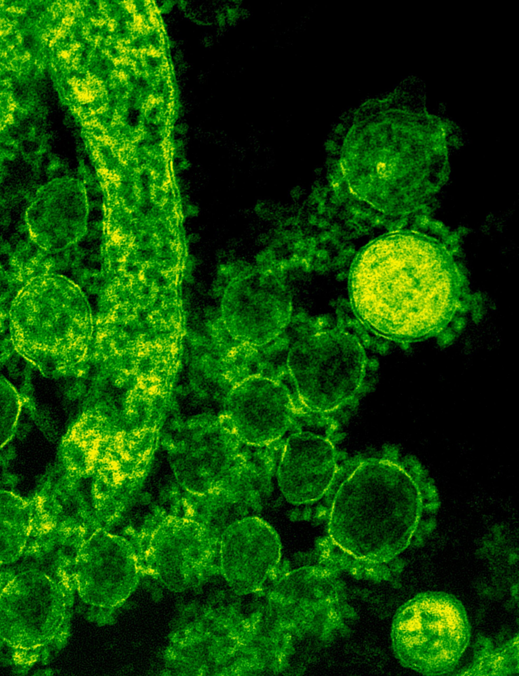
Header photo by National Cancer Institute on Unsplash
Quality control of HCS-HTS fluorescence imaging systems
In the landscape of high-content screening (HCS) and high-throughput screening (HTS) fluorescence imaging systems, precision and reliability take...
Precision Partners: Innopsys and Argolight on the InnoQuant Slide Scanners
In the intricate realm of pathology, drug discovery, and advanced research in brain function, cancer, and stem cells, the role of slide scanners has...
Lordil Microscopy announces the first quality control service using Argolight products.
Lordil Microscopy (lordil.fr) announces the first quality control service using Argolight products. This innovative service is offered through a...


